Our Services
1st Trimester Scan
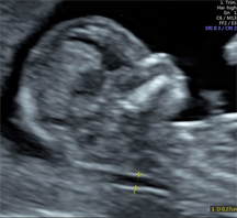
This scan is performed between 11 and 14 weeks gestation. In most cases it is performed transabdominally but it may sometimes be necessary to perform part of the examination transvaginally.
During this assessment, the pregnancy will be accurately dated and viability will be confirmed. The presence of multiple pregnancy will also be assessed for, and if confirmed, chorionicity will be determined - whether each fetus has their own placenta or if there is a single shared placenta, the latter posing higher risk to the pregnancy.
The anatomy of the fetus will be assessed for structural abnormalities and chromosomal markers that are visible in the 1st trimester. The nuchal translucency, nasal bone and facial profile will be assessed. This, in combination with a detailed maternal history and biochemical screening will be used to calculate the combined risk of the most common chromosomal abnormalities – Down’s Syndrome (Trisomy 21), Edward’s Syndrome (Trisomy 18) and Patau’s Syndrome (Trisomy 13).
The growth and development of the fetus will be assessed relative to gestational age. The placental circulation will be assessed via uterine artery Doppler sonography, and in combination with maternal history, blood pressure and biochemical screening, the risks of fetal growth restriction and pre-eclampsia will be calculated.
The risks of preterm delivery will be calculated by taking into account the maternal history and the measurement of the the cervical length during the assessment.
Patients will receive full counselling along with their results, and a management plan which will be formally communicated to their referring Obstetrican Gynaecologists. Those who are high-risk for chromosomal abnormalities including Down’s Syndrome will be offered further testing – invasive (Chorionic Villus Sampling/ Amniocentesis) or Non-Invasive Prenatal Testing (NIPT).